ReVISION
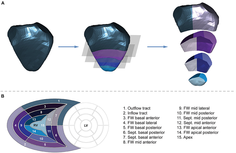
In heart diseases such as arrhythmogenic cardiomyopathy, pulmonary hypertension, tetralogy of Fallot and tricuspid regurgitation, early detection of right ventricular dysfunction leads to better outcomes. ReVISION divides the right ventricle into 15 segments and quantifies each segment's function so doctors can diagnose heart diseases earlier.
Today, right ventricular ejection fraction (RVEF) is a widely used measure of right ventricle (RV) performance. In many cases, however, a decline in global systolic function (measured by RVEF) occurs late in the progression of a patient's disease, sometimes too late. Doctors and patients who rely on RVEF to detect disease can miss the opportunity for early intervention and treatment.[1]


These diseases are often detectable in their early stages as they cause tell-tale changes in the work done by different segments of the RV. As different regions of the RV begin to be compromised by the early stages of the disease, other regions of the RV compensate to bear the load. Global systolic function, however, can often be unaffected. In pulmonary hypertension and in congenital heart diseases, for example, measuring RV function segment by segment can reveal adaptations to pressure and volume overload and, as a result, reveal the presence of disease at an early stage.[2]
Another example is in cases of arrhythmogenic cardiomyopathy. This is a heritable disease of the myocardium characterized by heart muscle loss and its replacement by fatty tissue, primarily in the right ventricle [3]. Because in the early stages of this disease the sections of lost muscle are small, they are very difficult to detect with conventional echocardiography. By measuring the function of different segments of the RV, however, the presence of these areas may be detected earlier as their effect on specific segments is typically measurable well before they would appear on a conventional echocardiographic analysis.

ReVISION divides the RV into 15 volumetric segments and quantifies corresponding EF and strain values to provide a meaningfully better diagnostic solution for the diseases mentioned above. Using ReVISION, the echocardiographer has a unique possibility to assess all of the surface of the RV, including such anatomically important parts as the RV outflow tract or the RV apex. Imaging and quantification of these parts are traditionally the most problematic to conventional assessments. Because ReVISION can measure RV function segment by segment, it can reveal diseases at a much earlier stage than other approaches can.
One characteristic that all heart diseases that impact the right ventricle have, is that patients whose diseases are detected early typically have better outcomes than those that do not.

Because so many of these diseases have subtle effects on the RV in their early stages, ReVISION, and its ability to measure RV function by segment, allows doctors and patients to chart a course to the best outcome possible.
To try ReVISION for yourself, click to see how ReVISION provides a comprehensive and quantified view of RV function.
Pricing Plan
Standard plan
$500 / month
-
User accounts that can run analyses and export data: up to 3
-
Supports 1 project with a single combined dataset
-
Supports export of analysis results in table format
-
ReVISION features enabled: all
-
Included raw 3D echo DICOM uploads per month: 50
-
Each additional raw 3D echo DICOM upload: $10 USD
-
TomTec 3D model uploads: Unlimited
-
Delivery: software as a service via
revision-research.arguscognitive.com -
7 day risk free trial
-
Included raw 3D echo DICOM uploads in the free trial: 10
-
Included TomTec 3D model uploads in the free trial: 10
References
[1] Surkova, Elena et al. “Contraction Patterns of the Right Ventricle Associated with Different Degrees of Left Ventricular Systolic Dysfunction.” Circulation. Cardiovascular imaging vol. 14,10 (2021): e012774. doi:10.1161/CIRCIMAGING.121.012774
[2] Bidviene J, Muraru D, Maffessanti F, Ereminiene E, Kovács A, Lakatos B, Vaskelyte JJ, Zaliunas R, Surkova E, Parati G, Badano LP. Regional shape, global function and mechanics in right ventricular volume and pressure overload conditions: a three-dimensional echocardiography study. Int J Cardiovasc Imaging. 2021 Apr;37(4):1289-1299. doi: 10.1007/s10554-020-02117-8. Epub 2021 Jan 3. PMID: 33389362; PMCID: PMC8026459.
[3] Malik N, Mukherjee M, Wu KC, Zimmerman SL, Zhan J, Calkins H, James CA, Gilotra NA, Sheikh FH, Tandri H, Kutty S, Hays AG. Multimodality Imaging in Arrhythmogenic Right Ventricular Cardiomyopathy. Circ Cardiovasc Imaging. 2022 Feb;15(2):e013725. doi: 10.1161/CIRCIMAGING.121.013725. Epub 2022 Feb 11. PMID: 35147040.