ReVISION
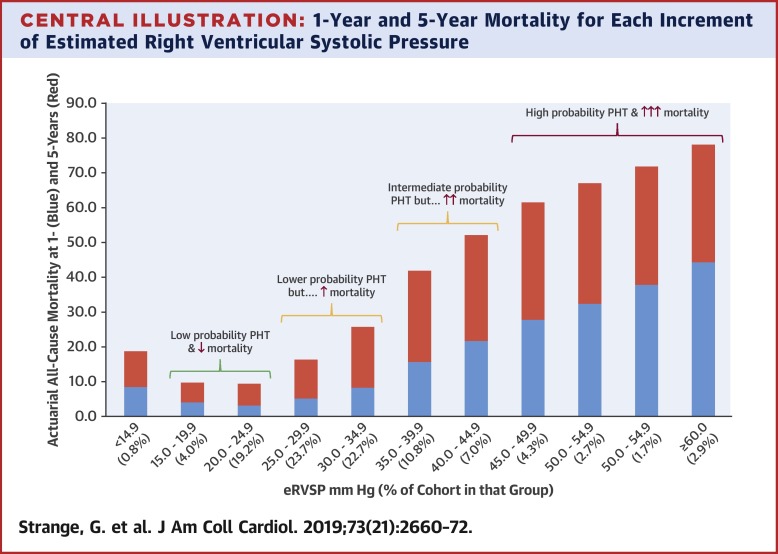
40% of patients with Pulmonary Hypertension die within 5 years of diagnosis. Others live for decades. ReVISION provides a quantitative, comprehensive view of right ventricle function, so doctors can help patients understand what PH will mean for them.
Nearly 80 million people worldwide, including up to 10% of elderly people, suffer from pulmonary hypertension (PH). PH often leads to right heart failure and premature death. 40% of patients diagnosed with PH die within 5 years of diagnosis, other patients can live for decades.

Patients with PH want to understand what the diagnosis means for them and for their quality of life. Because of the potential severity and the wide range of outcomes, giving an accurate prognosis depends on understanding how well, or poorly the patient's right ventricle is functioning.
Right ventricular (RV) function is the major factor that determines symptom burden, clinical outcomes, and survival among patients with PH. Until now, medical professionals have faced a difficult dilemma in evaluating right ventricle function.

Systems that give a comprehensive view of the right ventricle, such as 3D echocardiograms, do not provide quantitative measures of RV function that are sufficient to guide doctors to an accurate prognosis. Current quantitative measures do not provide a comprehensive picture of RV function because the RV's shape and function are complex.

Even the most recent data about the prognostic value of RV function relies on simple echocardiographic parameters and no established tool is available for detailed analysis of RV mechanics.
ReVISION, from Argus Cognitive, is a system that provides a deeper analysis of RV 3D echocardiographic data. ReVISION decomposes the global motion of the RV into three anatomically relevant components, along three independent axes.
ReVISION provides global and segmental functional parameters of the RV. Beyond established markers of ventricular mechanics (longitudinal and circumferential strain) the software quantifies RV wall motion directions and their impact on global and segmental volume changes.

ReVISION's comprehensive quantification of RV function has significant prognostic value. In particular, studies conducted with ReVISION have shown that the measurement of the anteroposterior component of RV motion (a capability unique to ReVISION) provides independent prognostic value.
ReVISION is suitable for use in everyday clinical practice in patients with PH and permits earlier detection of RV dysfunction and more granular risk stratification to support clinical decision-making.
To try ReVISION for yourself, click to see how ReVISION provides a comprehensive and quantified view of RV function.
Pricing Plan
Standard plan
$500 / month
-
User accounts that can run analyses and export data: up to 3
-
Supports 1 project with a single combined dataset
-
Supports export of analysis results in table format
-
ReVISION features enabled: all
-
Included raw 3D echo DICOM uploads per month: 50
-
Each additional raw 3D echo DICOM upload: $10 USD
-
TomTec 3D model uploads: Unlimited
-
Delivery: software as a service via
revision-research.arguscognitive.com -
7 day risk free trial
-
Included raw 3D echo DICOM uploads in the free trial: 10
-
Included TomTec 3D model uploads in the free trial: 10
References
Hassoun PM. Pulmonary Arterial Hypertension. N Engl J Med. 2021 Dec 16;385(25):2361-2376. doi: 10.1056/NEJMra2000348. PMID: 34910865.
Tokodi M, Staub L, Budai Á, Lakatos BK, Csákvári M, Suhai FI, Szabó L, Fábián A, Vágó H, Tősér Z, Merkely B, Kovács A. Partitioning the Right Ventricle Into 15 Segments and Decomposing Its Motion Using 3D Echocardiography-Based Models: The Updated ReVISION Method. Front Cardiovasc Med. 2021 Mar 4;8:622118. doi: 10.3389/fcvm.2021.622118. PMID: 33763458; PMCID: PMC7982839.
Bidviene J, Muraru D, Maffessanti F, Ereminiene E, Kovács A, Lakatos B, Vaskelyte JJ, Zaliunas R, Surkova E, Parati G, Badano LP. Regional shape, global function and mechanics in right ventricular volume and pressure overload conditions: a three-dimensional echocardiography study. Int J Cardiovasc Imaging. 2021 Apr;37(4):1289-1299. doi: 10.1007/s10554-020-02117-8. Epub 2021 Jan 3. PMID: 33389362; PMCID: PMC8026459.
Surkova E, Kovács A, Tokodi M, Lakatos BK, Merkely B, Muraru D, Ruocco A, Parati G, Badano LP. Contraction Patterns of the Right Ventricle Associated with Different Degrees of Left Ventricular Systolic Dysfunction. Circ Cardiovasc Imaging. 2021 Oct;14(10):e012774. doi: 10.1161/CIRCIMAGING.121.012774. Epub 2021 Sep 30. PMID: 34587749; PMCID: PMC8522626.
